
BikesGeek
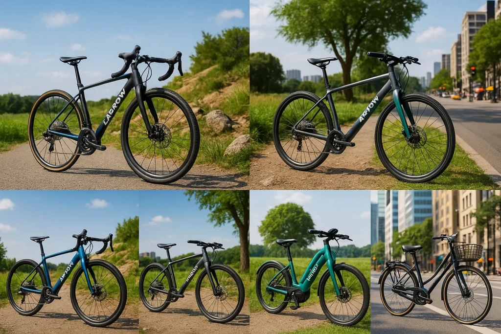
Why Zarrow Bikes Are the Best Choice for Every Rider
Cycling is a popular and sustainable way to stay fit, explore the outdoors, and reduce your carbon footprint. Over the past decade, the cycling industry has seen significant growth, with more people embracing cycling as their primary mode of transportation. Among the many brands on the market, Zarrow Bikes stands out for offering high-performance bicycles that meet the needs of different riders, from urban commuters to outdoor adventurers. With sleek designs, cutting-edge technology, and a commitment to sustainability, Zarrow Bikes provides the perfect balance of style, comfort, and reliability. In this article, we’ll explore why Zarrow Bikes should be your…

Zarrow Bikes: Changing the Way We Ride
Riding bikes is a great way to stay healthy, explore, and help the environment. Over time, the bike industry has worked hard to create bikes that are better, faster, and more fun to ride. Zarrow Bikes is a company that is making a big difference by designing high-quality bikes for all types of riders. Whether you ride to school, go on weekend adventures, or race professionally, Zarrow Bikes has a bike for you. Why You Should Choose Zarrow Bikes Zarrow Bikes is known for making some of the best bikes on the market. They have bikes for every kind of…

The Best Road Bikes for Beginners: A Comprehensive Guide
Finding the perfect road bike can be an exciting yet challenging task, especially for beginners. Whether you’re starting your cycling journey or looking to upgrade from a basic bike, it’s essential to choose a model that balances comfort, performance, and price. The best road bikes for beginners are those that offer ease of use, quality components, and a price point that doesn’t break the bank. In this guide, we’ll explore top recommendations, key features to consider, and helpful tips to make your decision easier. With so many options available, we’ve rounded up the most reliable bikes for newcomers to road…
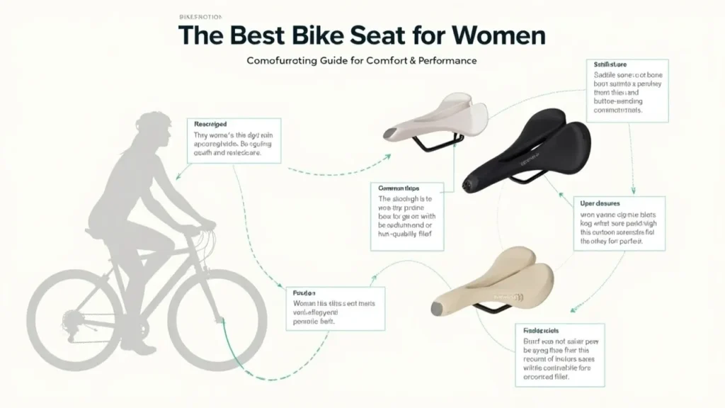
The Best Bike Seat for Women: A Comprehensive Guide for Comfort and Performance
Cycling has become an incredibly popular sport and recreational activity among women, and finding the perfect bike seat is crucial for making the experience comfortable and enjoyable. Whether you’re biking for fitness, commuting, or embarking on long-distance tours, the right seat can make all the difference in preventing discomfort, pain, and even injuries. This guide will walk you through everything you need to know to select the best bike seat for women in 2025. Why Choosing the Right Bike Seat for Women Matters Cycling is a fantastic way to stay fit and explore the outdoors, but it’s easy to overlook…

Best Long Distance Bikes 2025 - The Ultimate Guide
Long-distance cycling is more popular than ever, with more people exploring cycling as a means of travel, fitness, and adventure. As we approach 2025, the market for long-distance bikes is evolving with cutting-edge technologies, lightweight materials, and innovative designs. If you’re looking to ride further, faster, and more comfortably, choosing the best long distance bikes 2025 is crucial. In this guide, we’ll explore the top bikes for endurance, comfort, and performance, and help you choose the perfect ride for your next adventure. Why Long-Distance Biking is More Popular Than Ever Cycling offers numerous benefits—it’s an eco-friendly way to travel, an…

Best Bikes for Plus Size Women: A Complete Guide to Choosing the Right Ride
Introduction: Understanding the Need for Bikes for Plus Size Women Finding the right bike for plus size women is essential for comfort, safety, and enjoyment. Many women of larger sizes face challenges when searching for a bike that fits properly and can handle their weight. This is where specialized bikes for plus size women come in, designed to provide the support and durability needed for a comfortable ride. Cycling offers numerous benefits for women of all sizes. It promotes physical health, aids in weight loss, and enhances mental well-being. Plus, cycling is a low-impact activity that helps improve cardiovascular health…